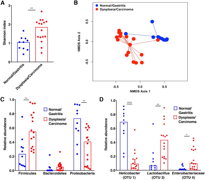
FIG 5.
The severity of histologic gastric injury following H. pylori infection significantly alters the diversity and structure of gastric mucosal microbiota, independent of host iron status. Gastric tissue from uninfected gerbils or gerbils infected with wild-type H. pylori strain 7.13 maintained on either iron-depleted or iron-replete diets was harvested 6 weeks postchallenge in linear strips, extending from the squamocolumnar junction to the proximal duodenum, and then homogenized. Microbial DNA was extracted from gastric tissue and subjected to 16S rRNA gene sequencing. (A) α-Diversity of the gastric microbiota was measured by Shannon diversity metric among H. pylori-infected gerbils maintained on either iron-depleted or iron-replete diets and stratified based on the severity of histologic injury. (B) β-Diversity of the gastric microbiota was measured by Yue and Clayton’s measure of dissimilarity and is shown in a nonmetric multidimensional scaling plot among H. pylori-infected gerbils maintained on either iron-depleted or iron-replete diets and stratified based on the severity of histologic injury. Normal/Gastritis (n = 10) versus Dysplasia/Carcinoma (n = 16), P < 0.001. (C) The relative abundance of phyla within the gastric microbiota was determined among H. pylori-infected gerbils maintained on either iron-depleted or iron-replete diets and stratified based on the severity of histologic injury. (D) The LDA effect size (LEfSe) algorithm was used to identify operational taxonomic units (OTUs) that were differentially abundant among H. pylori-infected gerbils maintained on either iron-depleted or iron-replete diets and stratified based on the severity of histologic injury. *, P, <0.01; **, P < 0.001; ****, P < 0.00001.

