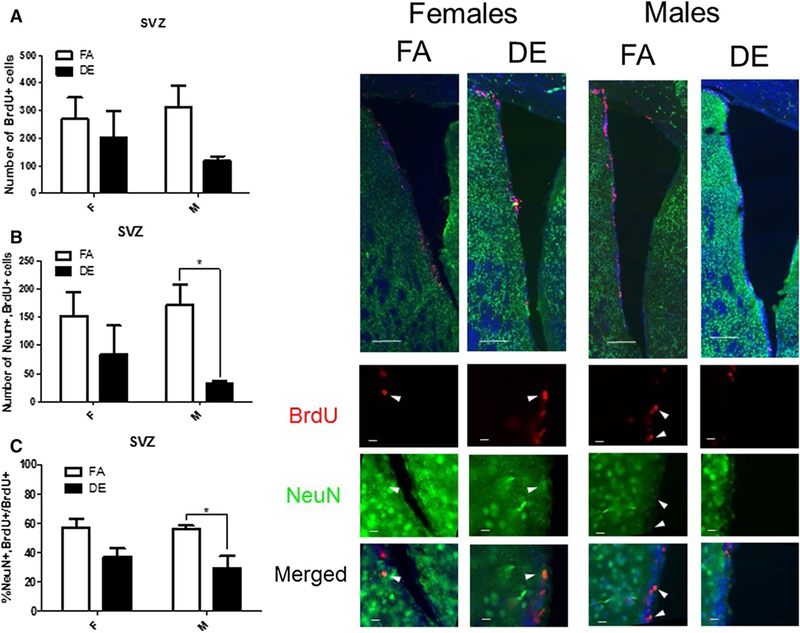
Fig. 3.

Effect of acute DE exposure on adult neurogenesis in the SVZ of mice. Eight-week-old male and female C57BL/6J mice were given five 100-mg/kg doses of BrdU at 2-h intervals. On the following day, they were exposed to DE (250 μg/m3) or FA for 6 h, and then sacrificed 21 days later. Images shown are representative micrographs of NeuN/BrdU immunohistochemistry in the SVZ of FA- and DE-exposed mice. Scale bars in montage images represent 100 microns, while scale bars in detail images represent 10 μm. The number of BrdU-stained cells (a) indicates surviving cells that been born since the beginning of the experiment. Double-stained NeuN/BrdU cells indicate surviving adult-born neurons, which are expressed both as a total number per brain region (b) and as a percentage of all BrdU-positive cells (c). Data shown represent the mean (± SE) with n = 3 per group. Significantly different from FA, *p < 0.05
