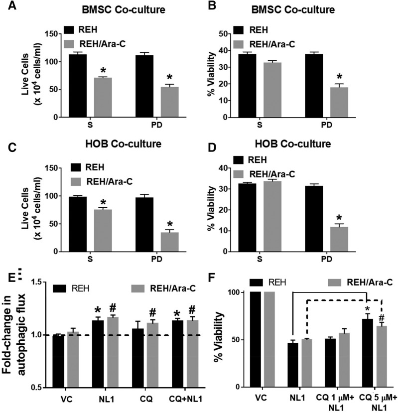
Fig. 6.
NL-1 induces cell death in coculture through an autophagy-dependent mechanism. REH and REH/Ara-C cells were grown in a coculture with either BMSCs or HOBs in a 24-well plate for 12 days. On day 9 in coculture, the cells were treated with 60 μM NL-1, chloroquine (CQ), or vehicle/DMSO. At the end of treatment, the total live cells in suspension (S) and the leukemic cells buried under the adherent layer (PD) were counted using the trypan blue dye exclusion method (A and C). The total cells and the live cell population were used to calculate the percentage of viability (B and D). *P < 0.05, when compared with REH cells. (E) REH and REH/Ara-C cells were plated in a 96-well plate and selected wells were pretreated for 1 hour with CQ. Following CQ treatment, the cells were treated with 60 μM NL-1. After 6 hours of treatment, the autophagic flux was measured. *P < 0.05, when compared with REH control, #P < 0.05, when compared with REH-Ara-C control group. (F) REH and REH/Ara-C cells were plated at 5 × 10⁴ cells per well in a 96-well plate and treated with indicated concentrations of CQ. Following 1 hour of CQ treatment the cells were exposed to 60 μM NL-1. After 72 hours of NL-1 treatment the cell viability was analyzed as described in Materials and Methods. *P < 0.05, when compared with NL-1 REH–treated group. #P < 0.05, when compared with NL-1 REH/Ara-C–treated group. All data are presented as mean ± S.E.M. and are representative of a study performed in triplicate and conducted three independent times.

