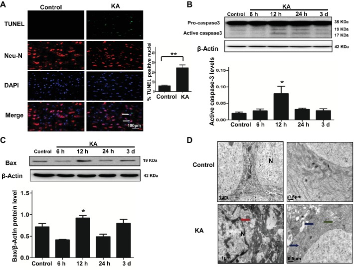
Figure 1.
Excitotoxic neuronal cell death is induced by addition of KA. (A) TUNEL assay showing apoptotic neurons in rat cortex samples. TUNEL-NeuN-positive cells are indicated by an arrow. Scale bar = 100 μm in all images. Western blot was performed for evaluation of active caspase-3 (B) and Bax (C) protein levels. *p < 0.05 vs. control (n = 6). (D) Neuron ultrastructure. Control group: Normal ultrastructure of neurons. KA group: Disrupted or shrunken nuclei (red arrow); swollen mitochondria (blue arrow), with dissolved and ruptured ridges; and dilated rough endoplasmic reticulum (RER) lumens (green narrow). KA, KA group; N, nucleus.

