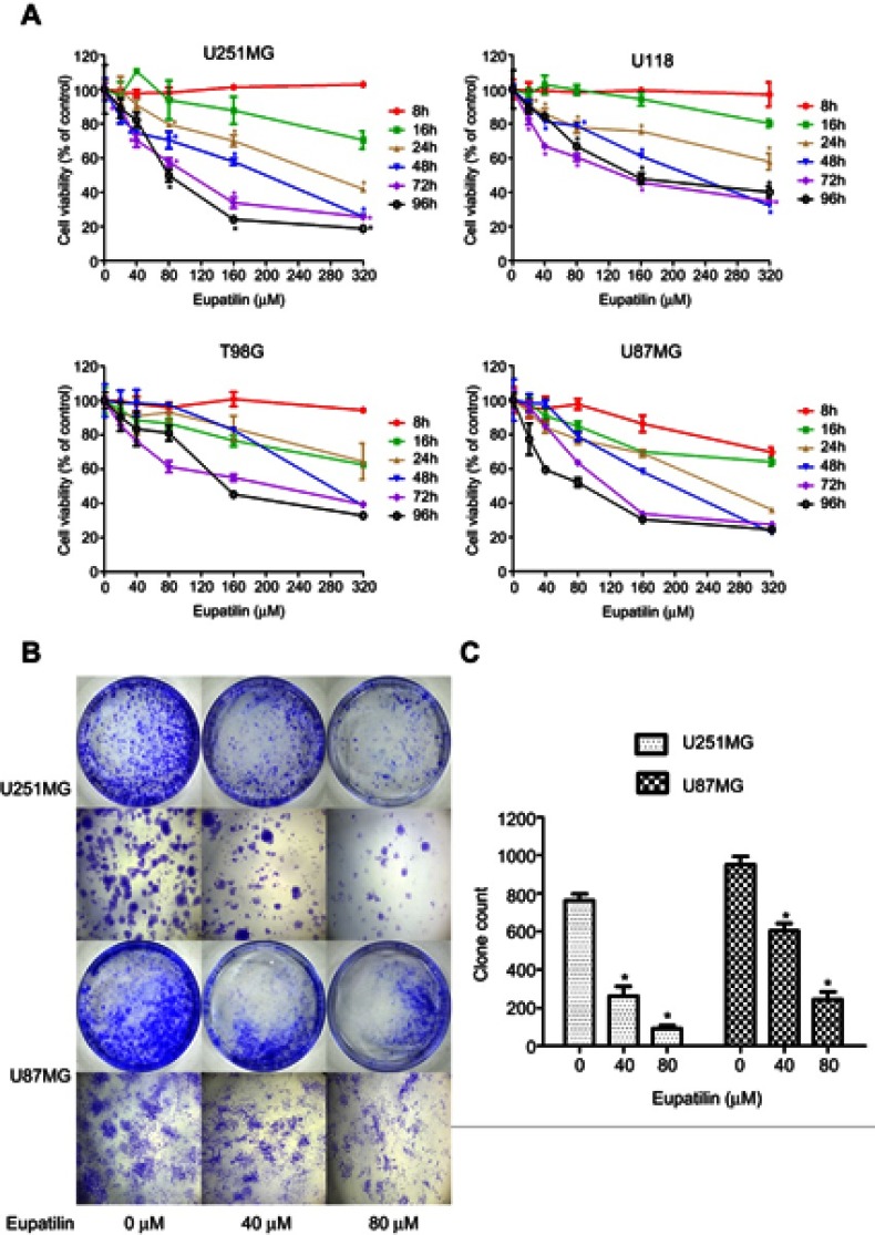
Figure 1.
Eupatilin inhibits viability and proliferation of glioma cells. (A) Four cell lines were treated with different concentrations (0, 20, 40, 80, 160, 320 μM) of eupatilin for different time points (8, 16, 24, 48, 72, 96 hrs), and the optical density values were measured at a wavelength of 450 nm. The viability rate of cells = (the OD values of treated groups/the OD values of the control group) × 100%. (B) U251MG and U87MG count 5000 cells each and were treated with 0, 40, 80 μM eupatilin for 15 days. The ability of eupatilin-treated tumor cells to form cell clones is significantly weaker than untreated cells. (C) Count the number of clones in four random fields in each dish. Data are expressed as the mean ± standard deviation of three independent experiments. *P<0.05 vs the control group. Each experiment was repeated three times.

