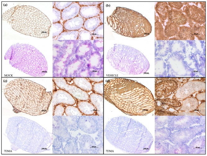
Figure 2.
Testicular levels of ZIKV antigens are reduced after 7DMA treatment as visualized by histopathological staining. Inoculation and treatment of AG129 mice was performed as described in Figure 3. The presence of ZIKV antigens (top panels in each quadrant) and inflammation (bottom panels in each quadrant) in the testis at day 10 pi is compared between mock-infected mice (a) and ZIKV-infected mice treated with vehicle (b) or 7DMA (c,d). The top two panels in each quadrant show antibody staining for the ZIKV envelope protein. The bottom two panels in each quadrant show hematoxylin and eosin staining. Panels on the left in each quadrant show a complete cross section of the testis. Panels on the right of each quadrant show a close up of the complete cross section of the same quadrant. The scale bars are 1000 µm and 200 µm for cross sections and close ups, respectively.

