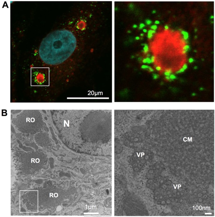
Figure 1.
Viral replication organelle in Japanese encephalitis virus (JEV)-infected cells. (A). fluorescent imaging of JEV replication organelle. At 48 h post-infection, JEV infected Vero cells were fixed, permeabilized, and stained with anti-nonstructural protein 4B (NS4B; red) and anti-double stranded RNA (dsRNA; Green) antibodies. Nuclear DNA was stained with DAPI (blue). Scale bar: 20 microns. (B). Viral replication organelle structure visualized using transmission electron microscopy (TEM). At 48 h post-infection, JEV infected Vero cells were fixed, ultra-thin section, and observed using TEM. N, nucleus; RO, viral replication organelle; CM, convoluted membrane; VP, vesicle packet. Scale bar = 1 micron (left) or 100 nm (right).

