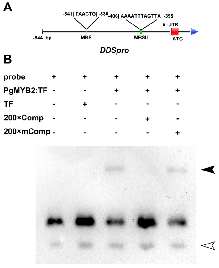
Figure 6.
Binding assay of PgMYB2 to the MBSII. (A) The diagram shows the relative location of the MBS and MBSII in the region of DDSpro. (B) The fusion protein of PgMYB2 binds to the MBSII of DDSpro. The reaction system from left to right: biotin-labeled probe (containing MBSII site); labeled probe and trigger factor as negative control (TF, a kind of chaperonin); labeled probe and PgMYB2: TF protein; labeled probe, PgMYB2: TF protein and 200× Comp (200 times unlabeled competitive probe); labeled probe, PgMYB2: TF protein and 200× mComp (200 times unlabeled competitive mutant probe, the MBSII site was mutated). The protein-probe complexes were indicated with a solid arrow and the free probes were indicated with a hollow arrow.

