Figure 1.
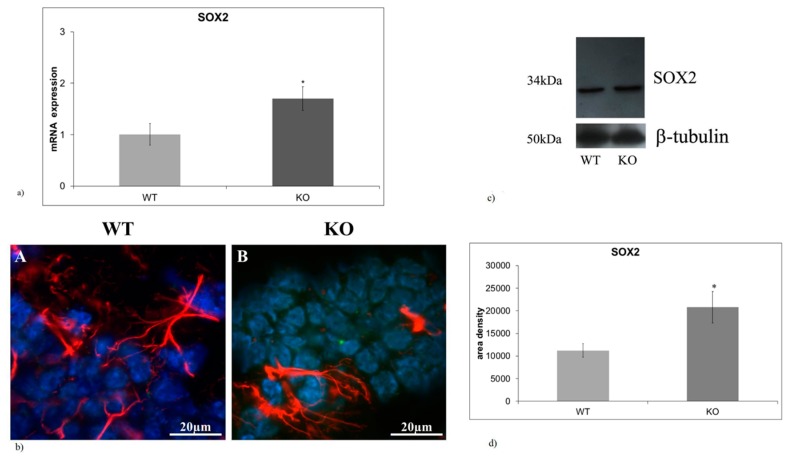
The absence of aSMase results in increase in SOX2 expression in hippocampal gyrus dentatus (a) Quantitative RT-PCR of SOX2 expression in the hippocampal GD of WT and aSMase-KO mice. (b) Immunofluorescences staining of SOX2 (green) and GFAP (red) in the hippocampal GD of WT e aSMase-KO mice. Immunofluorescence signal was analyzed as reported in materials and methods. The image shows merge between SOX2 and GFAP. Images were analyzed at 40X magnification. Scale bar = 20 μm. (c) Immunoblotting analysis of SOX2 expression in the hippocampal GD of WT e aSMase-KO mice. (d) Densitometry data of immunoblottingwere normalized by β-tubulin levels, expressed in arbitrary units. Data from (a,d) panels represent the mean ± SD of three independent experiments. ∗ p < 0.05 vs. WT mice. WT, wild type; KO, a-SMase-KO; GFAP, glial fibrillary acidic protein; SOX2, sex determining region Y-box 2.

