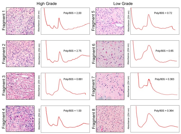
Figure 1.
Distinct regions of a human glioblastoma (GBM) tumor present very different histological characteristics and translational rates (original magnification 10×). The tumor was dissected in eight different fragments based on macroscopical appearance of the tissue. Histological characteristics of each fragment were evaluated by HE staining and classified in high- (numbered 1 to 4) or low-grade (numbered 5 to 8). Fragments were lysed and polysome profiles were obtained by separation using ultracentrifugation in a 7–47% sucrose gradient. As a measure of translation levels, the areas under the polysome and 80S peaks were determined and the ratios of polysome/80S peak were calculated.

