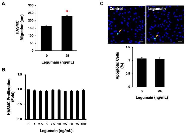
Figure 7.
Effects of legumain on the migration, proliferation, and apoptosis of HASMCs. (A) The effect of legumain on HASMC migration was determined in 10 cells per well using a BIOREVO BZ-9000 microscope. Three independent experiments were performed (n = 30). * p < 0.0001. Analyzed by unpaired Student’s t test. (B) The effect of legumain on HASMC proliferation was determined by WST-8 assay. Three independent experiments were performed (n = 3). (C) The effect of legumain on HASMC apoptosis was evaluated by detecting apoptotic cells (green) using a terminal deoxynucleotidyl transferase-mediated deoxyuridine triphosphate-biotin nick end-labeling (TUNEL) assay. Nuclei were co-stained with 6-diamidino-2-phenylindole (blue). The graph indicates the percentage of apoptotic cells in three independent experiments (n = 3). Scale bar = 100 μm.

