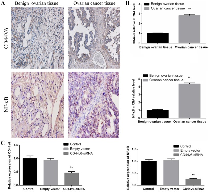
Figure 1.
CD44v6 and NF-κB expression in ovarian cancer tissues and an ovarian cancer cell line. (A) Immunohistochemical analysis of CD44v6 and NF-κB in benign ovarian tissue and malignant ovarian tumor tissue (×400 magnification). Brown represents positive expression of CD44v6 and NF-κB. CD44v6 was predominantly located on the cell membrane and NF-κB was primarily located in the nucleus of cancer tissue. (B) CD44v6 and NF-κB mRNA expression levels in benign ovarian tissue and malignant ovarian tumor tissue. **P<0.01 vs. benign ovarian tissue. (C) Relative mRNA expression levels of CD44v6 and NF-κB in different groups, including the control, empty vector and CD44v6-siRNA groups. No significant difference was identified between the control and empty vector groups. A significant difference was revealed between the control and CD44v6-siRNA groups. Data are presented as the mean ± standard deviation. **P<0.01 vs. control group. NF-κB, nuclear factor-κB; siRNA, small interfering RNA; CD44v6, cluster of differentiation variant 6.

