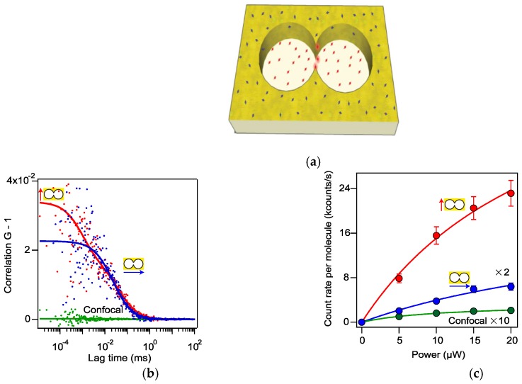
Figure 5.
(a) Sketch of a double nanohole under an incident light polarized along with the two tips. (b) Fluorescence correlation spectroscopies for with double nanohole using incident light polarized parallel (red color) and perpendicular (blue color) to the apex region, and with a confocal microscope (green color). Dotted data is the measurement result and the solid curve is the corresponding fitting result for each case. (c) Comparison in brightness of fluorescence among these three cases at different incident power. Adapted from [82], with permission © 2015 Springer Nature Publishing A.G.

