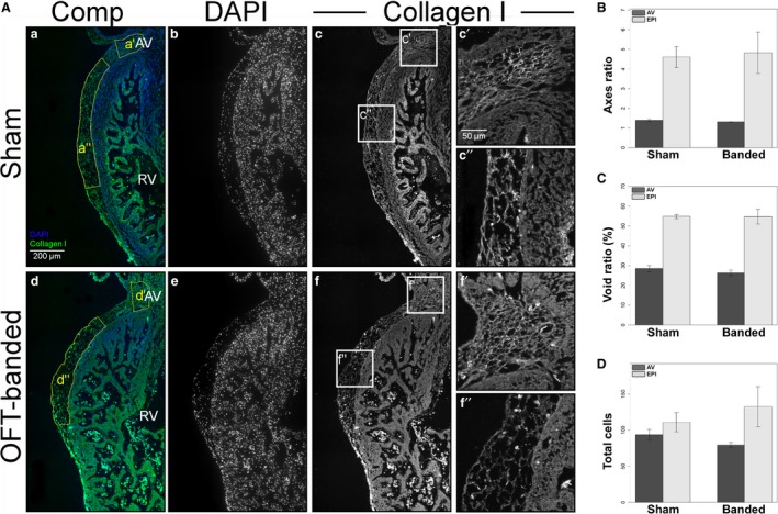
Figure 4.

Normal collagen I expression, morphology and cell counts in HH29 hearts. (A) Hearts were stained for DAPI (b,e) and collagen I (c,f, boxes c’,c”,f’,f”). The region of interest chosen for the statistical analysis is denoted by the yellow boxes (a’,a”,d’,d”) in composite images (a,d). Collagen I morphology can be seen in detail around the atrioventricular (AV) canal (c’,f’) and above the right ventricle (c”,f”). RV, right ventricle. Scale bar: 200 μm (b–f). Scale bars: 50 μm (c’,c”, f’,f”). (B) There was no significant difference in the axes ratio of AV canal and right ventricle epicardium (EPI) region. Error bars denote SEM. (C) There was no significant difference in the void ratio of the AV canal and right ventricle epicardium region (EPI) (collagen I area/total area*100). Error bars denote SEM. (D) There was no significant difference in the cell counts of the AV canal and right ventricle epicardium region (EPI). Error bars denote SEM.
