Figure 7.

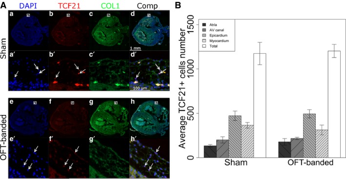
Numbers of TCF21+ cells are unaltered in HH35 hearts. (A) Hearts stained for DAPI (a,e), TCF21 (b,f) and collagen I (c,g). A composite can also be seen (d,h). Zoomed sections (a’,b’,d’,e’,f’,h’) show TCF21+ cells (arrows) as well as their collagen I surroundings (c’,g’). Scale bars: 1 mm (a,b‐h), 100 μm (a’,b’–h’). (B) No significant difference was found concerning the numbers of TCF21+ cells in the different heart regions between sham and OFT‐banded hearts. Error bars denote SEM. AV, atrioventricular.
