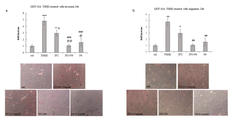
Figure 5.
Effect of 5FU, 5FU+F6, and F6 on invasion (a) and migration (b) in MCF-10A cells. Transwell assays were performed in MCF-10A cells untreated (ctrl), and pre-treated with TGF-β1 for 5 days. TGF-β1 stimulated MCF-10A cells were then treated with 5FU, 5FU+F6, and F6 for 24 h. Values, expressed as fold increase of control value considered as 1, are means of three independent experiments performed in duplicate, with SD represented by vertical bars. * p < 0.05; ** p < 0.01; *** p < 0.001 versus ctrl; # p < 0.05; ## p < 0.01; ### p < 0.001 versus TGF-β1; @ p < 0.05; @@ p < 0.01 versus 5FU by ANOVA followed by Bonferroni post-test. Images were obtained by optical microscopy, with 100× magnification.

