
Figure 1.
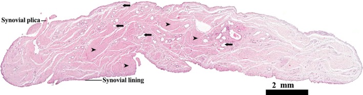
Transverse profile of the LHF mid‐way along its length. H&E stain. The full profile of the ligament is lined in synovial membrane, sections through synovial plicae are also evident. Scattered blood vessels (arrows) are visible both in the sub‐synovial layer and connective tissue of the ligament proper (arrowheads).