
Figure 2.
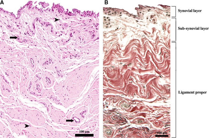
Tissue layers of the LHF. (A) H&E stain. Cuboidal synovial cells are visible on the surface (synovial layer); fibroblasts (arrowheads) and blood vessels (arrows) are present both in the sub‐synovial tissue and ligament proper. (B) VVG stain. Collagen fibres are stained in red and elastin in black.