
Figure 3.
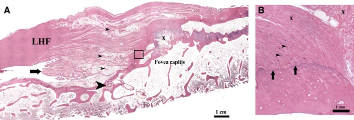
Histology of the LHF at its foveal attachment (H&E stain). (A) Ligamental blood vessels near the fovea capitis (small arrowheads). Patent vessels seen entering the foveal floor (large arrowhead). Section shows synovial plicae (arrow) and a prominent osteophyte (x). (B) Magnified view of the boxed area in (A): The enthesis shows a transition of collagen bundles (x) to fibrocartilage and to bone tissue at the foveal insertion. Chondrocytes are visible in the fibrocartilage (arrowheads). The tidemark (arrows) separates the fibrocartilage from bone.