
Figure 3.
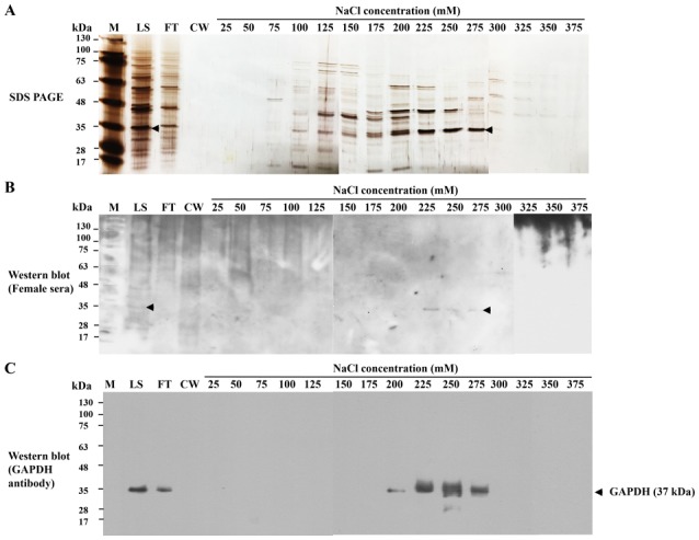
Purification of HeLa-GAPDH from HeLa cell cytosolic proteins by cation-exchange chromatography. (A) HeLa cell cytosolic proteins were separated by cation-exchange chromartography. The proteins were separated by SDS-PAGE and visualized with silver staining. (B) Western blot analysis of proteins from cation-exchange chromatography using healthy donor sera. (C) Western blot analysis where HeLa-GAPDH was detected in fractions from cation-exchange chromatography using anti-human GAPDH polyclonal antibody. HeLa-GAPDH is indicated by arrowheads. M, protein markers; LS, loading sample; FT, flow-through; CW, column wash.