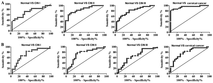
Figure 6.

ROC curves for discriminating CIN I, CIN II, CIN III and cervical cancer from healthy specimens. (A) ROC curves for discriminating cervical lesions from normal cytology based on the anti-HeLa-GAPDH IgG levels observed in Fig. 4A. (B) ROC curves for discriminating cervical lesions from normal cytology based on the anti-HeLa-GAPDH IgM level observed in Fig. 4B. ROC, receiver operating characteristic; CIN, cervical intraepithelial neoplasia.
