Figure 3.

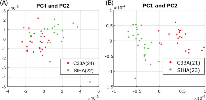
Standard (A) and modulated (B) spectral datapoints represented as PC1/2 weightings for C33A (HPV negative carcinoma) and SiHa (HPV‐16 carcinoma) cells. The number of cells used for each comparison are indicated in the legend

Standard (A) and modulated (B) spectral datapoints represented as PC1/2 weightings for C33A (HPV negative carcinoma) and SiHa (HPV‐16 carcinoma) cells. The number of cells used for each comparison are indicated in the legend