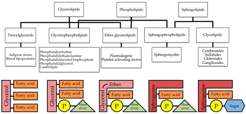
Figure 1.
Several forms of triglycerides and phospholipids found in the cell. The left-most scheme is a triglyceride formed by a three-carbon glycerol backbone with a FA tail bound to each carbon. Second left-most scheme is a phospholipid bearing two FA tails with the third carbon of glycerol bound to a phosphate group. The middle scheme is an ether glycerolipid, who shares with the two previous structures the three-carbon glycerol backbone and is slightly modified with an ether group. In second right-most scheme, a sphingosine is shown as backbone instead of glycerol and first fatty acid tail of glycerol is modified slightly. This is a sphingophospholipid since it bears a phosphate group, it is called sphingomyelin. Right-most scheme shows the scheme of sphingolipids called glycolipids, since the phosphate group is replaced with a sugar (carbohydrate group).

