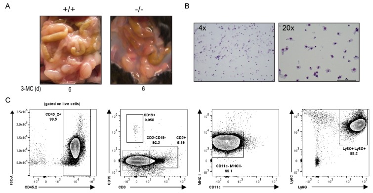
Figure 3.
Wright Giemsa stain and flow cytometry analysis of the chylous ascites collected from the peritoneum of 3MC-treated mice on day 6. (A) Accumulation of fluid observed in 3MC-treated WT and Tiparp−/− mice. (B) Cellular bodies present in the ascitic fluid. (C) To phenotype the cellular infiltrate, flow cytometry was used and identified the populations to be predominantly neutrophilic. Representative plot of four ascites samples showing a greater neutrophil population (Ly6C+, Ly6G+). Peritoneal fluid was stained directly. Almost all cells in fluid are hematopoietic (CD45.2+) and the majority are innate cell types (CD3- and CD19-). Of these innate cell types, they are not antigen-presenting cells (CD11c- and MHC II-).

