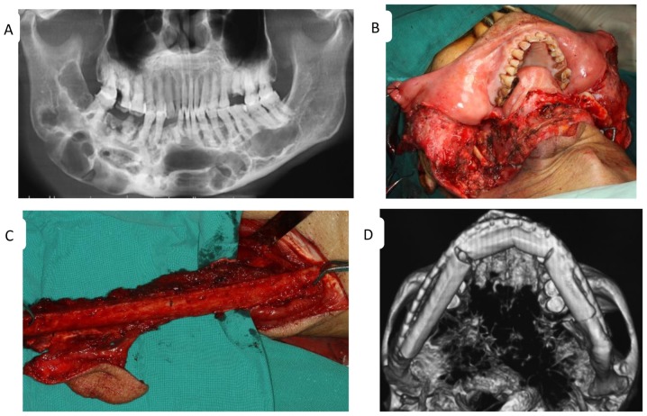
Figure 1.
Features of odontogenic keratocyst determined the treatment regimen. (A) A preoperative panoramic radiograph of a large lesion with multiple radiolucencies extending from the left third molar to the contralateral ascending branch of the mandible. Intraoperative views of (B) a segmental mandibulectomy and (C) the fibula flap. (D) An image of the reconstructed mandible following surgery.

