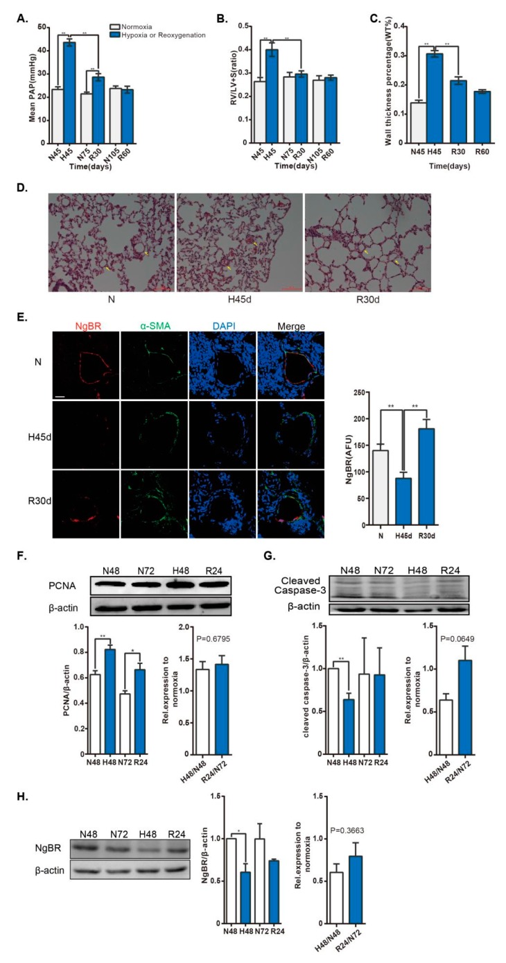
Figure 1.
NgBR expression is downregulated in the pulmonary artery of HPH rat model and in vascular smooth muscle cells (VSMCs) exposed to hypoxia. Rats were housed in hypobaric hypoxia (10.0% O2) for 45 days, and then exposed to reoxygenation at different times. Pulmonary vascular remodeling was evaluated by measuring the (A) mPAP and (B) weight ratio of RV/ (LV + S). (C,D) Histological images of distal pulmonary arteries (arrows)stained with hematoxylin and eosin. Scale bar = 100 µm. The percent medial wall thickness was analyzed using an Olympus IX-70 microscope. n = 44–50 pulmonary arteries (35–100 µm) from three animals/group. (E) The small pulmonary arteries (30–140 µm) were co-stained with α-smooth muscle actin (α-SMA; green) and NgBR (red). Nuclei were stained with DAPI (blue). Results were calculated as a relative arbitrary immunofluorescence unit (AFU) using FV10-ASW3.1 software. n = 38–54 pulmonary arteries from three animals/group. Scale bar = 30 µm. The VSMC cell line, A10, was exposed to 4% O2 for 48 h, followed by exposure to 21% O2 for 24 h. (F,G) Cell proliferation was evaluated by determining PCNA expression. n = 4. Apoptosis was detected by analyzing cleaved-capase-3 expression. n = 3. ** p < 0.01 H48 versus N48, R24 versus N72. (H) NgBR expression was normalized to β-actin. n = 3. * p < 0.05.

