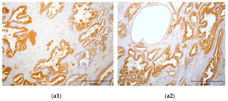
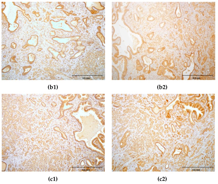
Figure 6.
(a1,b1,c1) Immunohistochemical staining of three different hPSA tissue samples with standard monoclonal antibody ER-PR8 (dilution 1:100). (a2,b2,c2) Immunohistochemical staining of three different hPSA tissue samples with biotinylated antisense peptide AVRDKVG (dilution 1:100). The black line in the lower right corner denotes the scale bar (0.5 mm).


