Figure 1.
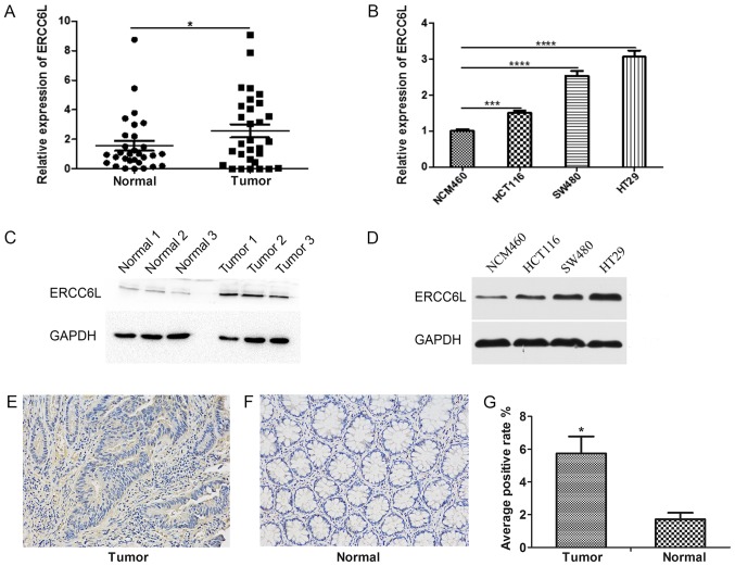
Analysis of ERCC6L expression in human CRC tissues and cell lines. (A) Reverse transcription-quantitative polymerase chain reaction was used to detect the relative expression levels of ERCC6L in 30 matched pairs of human CRC and noncancerous tissues. *P<0.05. (B) Relative expression of ERCC6L in three CRC cell lines (HCT116, SW480 and HT29) and one normal colonic mucosal cell line (NCM460). ***P<0.001 and ****P<0.0001. Expression level of ERCC6L in (C) three matched pairs of CRC and adjacent noncancerous tissues and (D) three CRC cell lines and one normal colonic mucosal cell line as detected by western blotting. GAPDH was used as an internal control. Representative images of IHC staining of ERCC6L in (E) a CRC tissue sample and (F) an adjacent normal tissue sample. Magnification, ×200. (G) IHC revealed a significant upregulation of ERCC6L protein in tumor samples compared with normal samples. *P<0.05 vs. normal. ERCC6L, excision repair cross-complementation group 6 like; CRC, colorectal cancer; IHC, immunohistochemistry.

