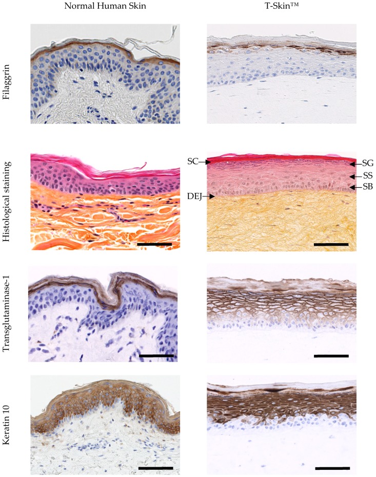
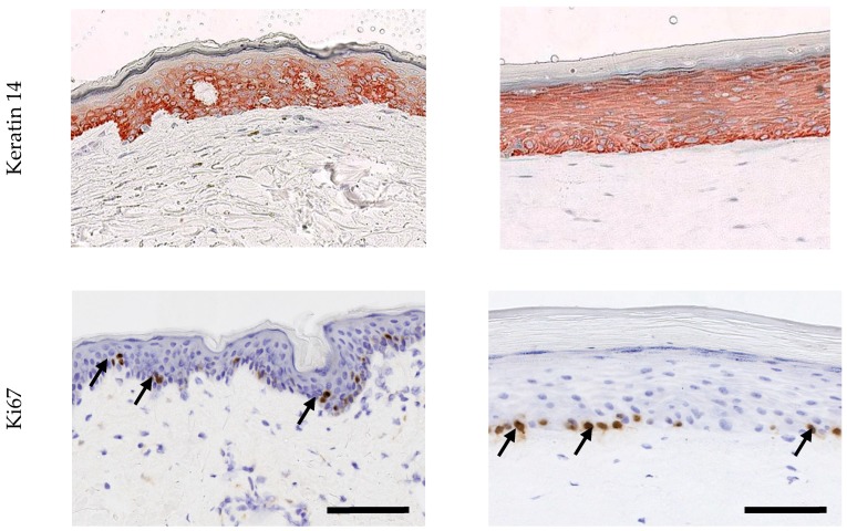
Figure 2.
Characterization of the epidermis from normal human skin (NHS) and T-Skin™ by histology and immunohistochemistry analysis on paraffin sections. Localization on histological stained images of the stratum corneum (SC), stratum granulosum (SG), stratum spinosum (SS), stratum basal (SB) layers of the epidermis and the dermo–epidermal junction (DEJ). For histological staining, the scale bar represents 100 μm. All other bars are 100 μm. Arrows indicate positive cells for Ki67 expression


