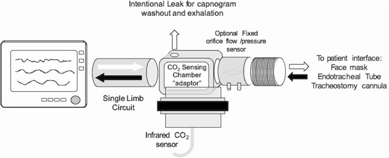
Fig. 1.
Experimental setup: single-limb circuit configuration. The experimental setup used to validate the end-tidal dilution method (etCO2D) to estimate auto-PEEP. See text for detailed explanations of the components. In this configuration, the exhaled air (black arrow) is always confronted with the continuous flow/pressure of fresh gas generated by the ventilator (white arrow). Whenever the end-expiratory pressure of the animal exceeds the pressure/flow coming from the ventilator, a complete normal capnogram is obtained from the CO2 sensor. If, however, the expiratory pressure of the ventilator exceeds the end-expiratory pressure of the patient, the capnogram will be diluted. In this evaluation, different levels of intrinsic PEEP were created and detected by stepwise increasing the level of end-expiratory pressure from the baseline value of 2 cmH2O in 1 and 0.2 cmH2O steps until de first dilution of the end-tidal portion of the capnogram was diluted. This pressure corresponded to the level of PEEPi

