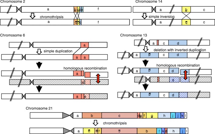
Fig. 2.
Schematic of predicted mutagenic processes for each rearrangement. Rearranged DNA fragments are highlighted in different colors. Underlined letters depict inverted DNA fragments. Striped boxes indicate non-sister chromatids in the father. Red arrows denote physiological homologous recombination during meiosis. This figure is not drawn to scale

