Fig. 1.
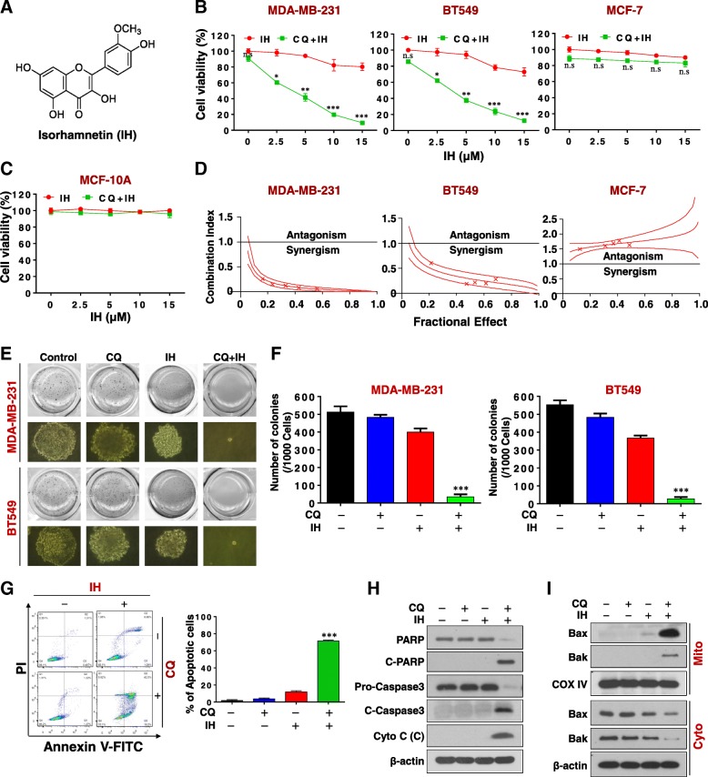
CQ dramatically potentiates IH-mediated inhibition of cell proliferation and the induction of apoptosis in TNBC cells. a The chemical structure of isorhamnetin (IH). b and c MDA-MB-231, BT549, MCF-7, and MCF-10A cells were treated with various concentrations of IH in the presence or absence of 20 μM CQ for 48 h, and MTT assays were performed to assess cell proliferation—mean ± SD for three independent experiments, ns, not significant, *P < 0.05, **P < 0.01 or ***P < 0.001 compared with IH. d The combination index (CI) values for each fraction affected were determined using commercially-available software (Calcusyn, Biosoft). CI values less than 1.0 correspond to synergistic interactions. e and f Colony formation was detected using a soft agar assay in MDA-MB-231 and BT549 cells (mean ± SD for three independent experiments, ***P < 0.001 compared with control). g-i MDA-MB-231 cells were combination treated with CQ (20 μM) and IH (10 μM) for 48 h. Apoptosis was determined by Annexin V-FITC/PI staining and flow cytometry (mean ± SD for 3 independent experiments; ***P < 0.001 compared with control or CQ and IH treatment alone). The total cellular extract, cytosol and mitochondrial fractions were prepared and subjected to western blot using antibodies against total PRAP, C-PARP, pro-Caspase 3, cleaved caspase-3, cytochrome c (Cyto C), Bak, and Bax. β-actin and COX IV were used as loading controls

