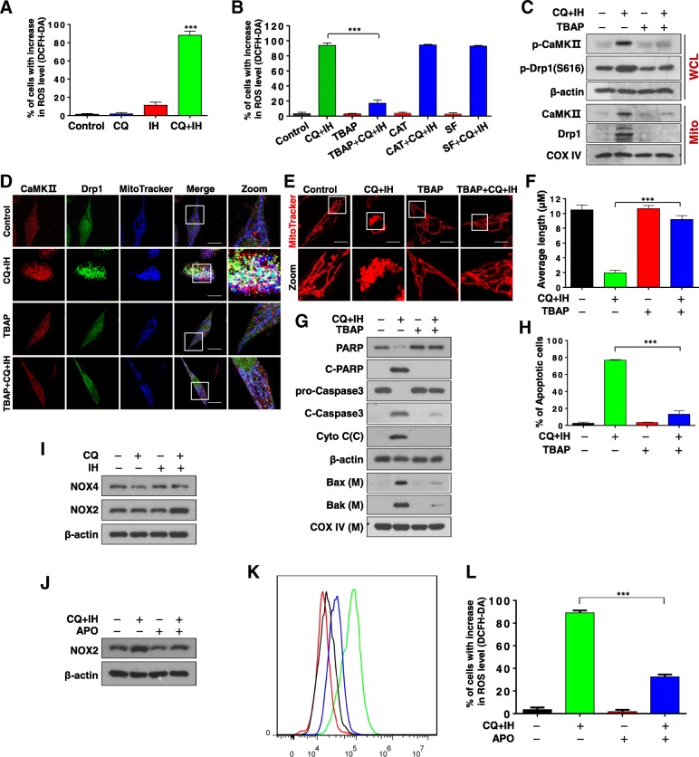
Fig. 5.
Effects of antioxidants on CQ/IH-induced ROS generation, mitochondrial fission, apoptosis, and cell signaling proteins. a MDA-MB-231 cells were treated with CQ (20 μM) in the presence or absence of IH (10 μM) for 6 h. Cells were stained with DCFHDA, and ROS production was analyzed by flow cytometry as described in Materials and Methods (mean ± SD for three independent experiments; ***P < 0.001 compared with control or CQ and IH treatment alone). b Cells were pretreated with antioxidants including TBAP (200 μM), catalase (5000 U/ml), and sodium formate (SF, 2 mM) for 1 h, followed by combined treatment with CQ/IH, after which ells were stained with DCFHDA; ROS production was then analyzed by flow cytometry (mean ± SD, ***P < 0.001). For C–H, cells were pretreated with TBAP, followed by the CQ/IH combination. c WCL and Mito were prepared and subjected to Western blot using antibodies against p-CaMKII (T286), p-Drp1 (S616), CaMKII, and Drp1. d The colocalization of CaMKII (red), Drp1 (green), and MitoTracker (blue) was examined by confocal microscopy. Scale bars: 10 μm. e Mitochondrial morphology was observed by MitoTracker Red CMXRos staining and confocal microscopy. Scale bars: 10 μm. f Mitochondrial length was measured with ImageJ software. Fifty cells of three independent experiments (mean ± SD, ***P < 0.001). g WCL, Cyto, and Mito were prepared and subjected to Western blot using antibodies against total PRAP, C-PARP, pro-Caspase 3, C-Caspase 3, cytochrome c, Bak, and Bax. h Apoptosis was detected by flow cytometry analysis. The values represent the mean ± SD for three separate experiments (mean ± SD, ***P < 0.001). i MDA-MB-231 cells were treated with CQ (20 μM) in the presence or absence of IH (10 μM) for 48 h. WCL were prepared and subjected to Western blot analysis using antibodies against NOX4 and NOX2, β-actin being used as a loading control. j Cells were pretreated with APO (100 μM) for 2 h, followed by the combination of CQ/IH. WCL were prepared and subjected to Western blot analysis using antibodies against NOX2, β-actin being used as a loading control. k and l Cells were pretreated with APO (100 μM) for 2 h, followed by the combination of CQ/IH for 6 h. Cells were stained with DCFHDA and ROS production was analyzed by flow cytometry. (mean ± SD for three independent experiments; ***P < 0.001)

