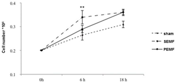
Figure 1.
Numbers of human gingival fibroblast (hGF) cells following exposure to two types of electromagnetic field (EMF). The hGF cell numbers were calculated after 6 and 18 h exposure to a sham, a sinusoidal electromagnetic field (SEMF), and a pulsed electromagnetic field (PEMF). The means ± standard deviation (SD) of the change from baseline (0 h) over time are reported in the graph. Experiments were carried out in duplicate, and hGFs derived from the 11 recruited patients, were analyzed. ** p-Value < 0.01 SEMF vs. sham.

