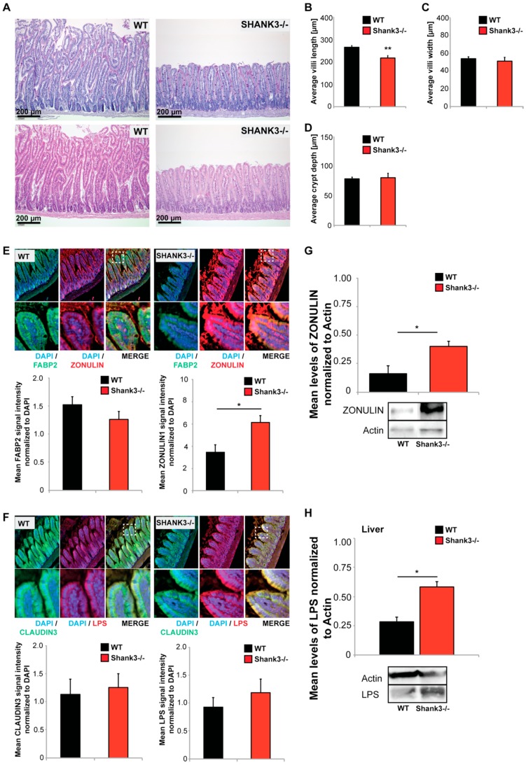
Figure 2.
Altered gut morphology in Shank3αβ knock-out (KO) mice. (A–D) Histological evaluation of GI tract from wild type and Shank3αβ KO mice. (A) Longitudinal cross sections of Shank3αβ KO mice and wild type (WT) mice were stained with hematoxylin/eosin (HE) (upper panels) and periodic acid schiff (PAS) reaction (lower panels). Exemplary images are shown. (B–D) Morphological analysis of (B) villi length and (C) width, and (D) crypt depth reveals a significantly decreased villi length (Mann-Whitney U-test, p = 0.009; n = 5 animals per group) but not width (p = 0.534), and normal crypt depth (p = 0.983) in Shank3αβ KO mice. (E,F) Immunohistochemistry was performed on 5 mice per group and 5 optic fields of view each from 3 sections per mouse were analyzed. (E) A slight but non-significant decrease in FABP2 signal intensity was observed in Shank3αβ KO mice compared to wild types (left panel). Significantly higher ZONULIN-1 levels were found in Shank3αβ KO mice (right panel) (t-test, p = 0.0413). (F) The levels of CLAUDIN3 and lipopolysaccharide (LPS) were not significantly different between Shank3αβ KO mice and wild types in gut epithelium. (G) Significantly higher ZONULIN-1 levels in Shank3αβ KO mice were confirmed by western blotting using gut epithelium protein lysate (t-test, p = 0.0434, n = 3 per group). (H) Protein lysate from liver tissue from WT and Shank3αβ KO mice (n = 3 per group) were analyzed for E. coli LPS levels using Western Blotting. The results show significantly higher LPS levels in the liver of Shank3αβ β KO mice (t-test, p = 0.0452). * p < 0.05, ** p < 0.01.

