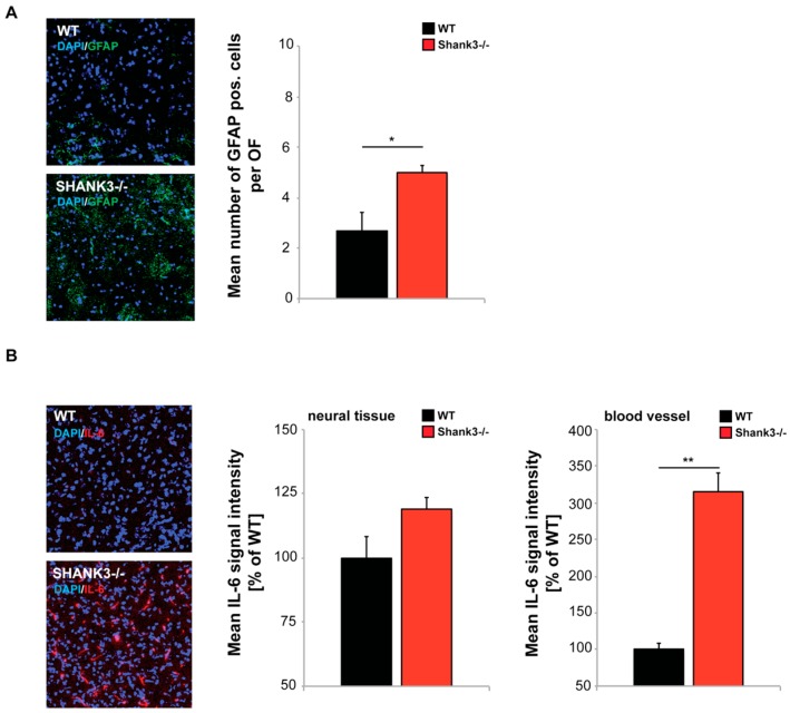
Figure 4.
Confocal microscopy images with same acquisition time taken from frontal cortex of brain sections from WT and Shank3αβ KO mice (n = 3 animals per group) were used to assess the number of activated astrocytes labeled by Glial fibrillary acidic protein (GFAP), and IL-6 levels in neural tissue and blood vessels. DAPI staining was used to visualize cell nuclei. (A) Optic fields (OF) of view were analyzed and the number of GFAP positive cells per OF measured. The results show a significantly higher number of activated astrocytes in Shank3αβ KO mice (t-test, p = 0.0407). (B) The immunofluorescence of IL-6 was slightly higher in Shank3αβ KO mice compared to WT (t-test, p = 0.1164) in neural tissue and significantly higher in blood vessels (t-test, p = 0.0014). * p < 0.05, ** p < 0.01.

