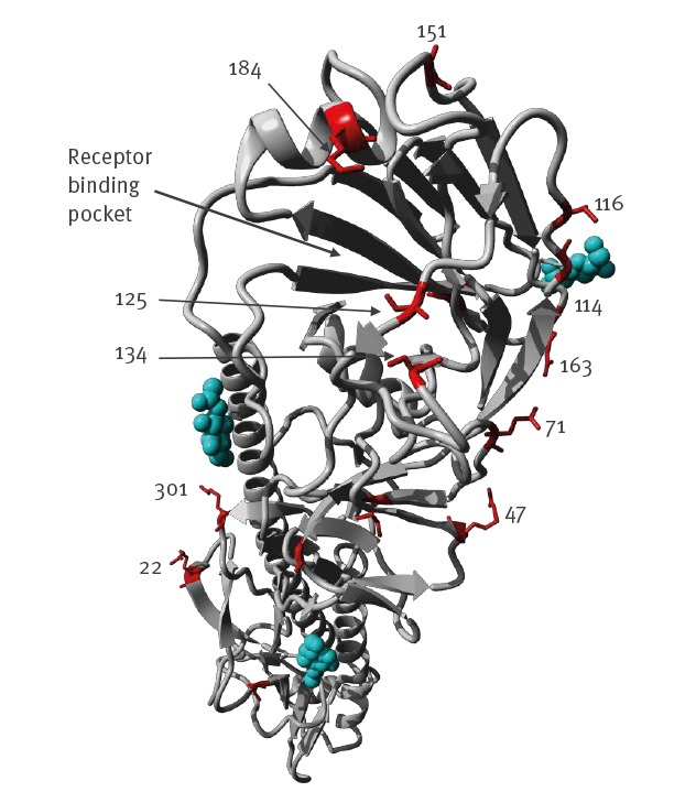
Figure 5.
Structural view of mutations in the haemagglutinin 1 viral sequences recovered from a highly pathogenic avian influenza A(H7N9) infected patient and from environmental samples collected nearby, compared to candidate vaccine strain A/Guangdong/17SF003/2016(H7N9), Inner Mongolia Autonomous region, China, April 2019
Residues at positions of mutations listed in Table 1 are shown as red sticks in the crystal structure of H7N9 HA (PDB: 4KOL, using YASARA). Glycosylation as seen in original structure is shown as cyan balls.