Figure 3.

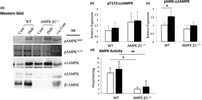
AMPK phosphorylation in kidneys from diabetic and non‐diabetic WT and AMPK β1−/− mice 28 days after induction of diabetes. A Lysates were immunoprecipitated with a mixture of anti‐α1 and anti‐α2 AMPK antibodies, and then blotted and probed with antibodies against pT172, pS485, both α subunits of AMPK, and the β1 subunit. B, Densitometric analysis of Western blots showing relative expression of pαT172. WT control n = 3, WT diabetic n = 5, AMPK β1−/− control n = 3, AMPK β1−/− diabetic n = 5. C, Densitometric analysis of Western blots showing relative expression of pαS485. n = 3‐5. D, AMPK activity assay in kidneys from diabetic and non‐diabetic WT and AMPK β1−/− mice 28 days after induction of diabetes. Kidney lysates were immunoprecipitated with both α1 and α2 AMPK antibodies and the immunoprecipitates assayed by SAMS assay. (B) Adiponectin: WT control n = 3, WT diabetic n = 6, AMPK β1−/− control n = 3, AMPK β1−/− diabetic n = 5. *P < 0.05, **P < 0.01. Non‐diabetic (white bars), diabetic (black bars)
