Figure 8.

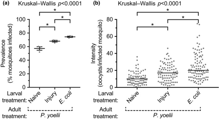
Prevalence and intensity of malaria infection in adult female mosquitoes following various larval treatments. Larvae were left unmanipulated (naïve), injured, or infected with E. coli. Five days after eclosion, the adults were blood fed on mice infected with Plasmodium yoelii, and the prevalence (a; percent of mosquitoes with oocysts; horizontal line and whiskers denote the mean and the standard error of the mean, respectively) and intensity (b; circles indicate the number of oocysts in infected mosquitoes and the horizontal line marks the median) of infection was measured 7–9 days later. Asterisks denote significant differences (Dunn's p < 0.05) between the groups
