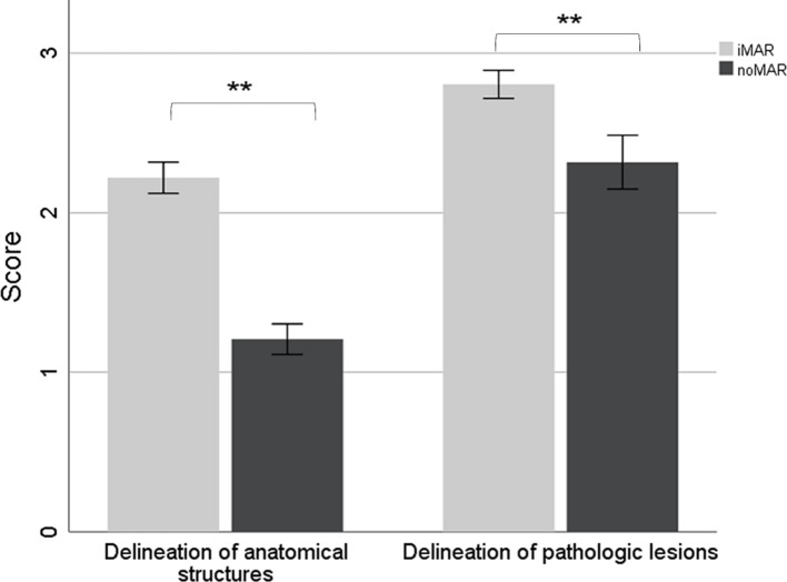
Figure 6.
3-point Likert scale showing the effect of iMAR on the subjective image quality rated by two independent radiologists. Severity of artifacts in iMAR corrected and noMAR CT for the analysis of anatomical structures (left side; score ±95% CI) and delineation of pathologic lesions in iMAR corrected and noMAR CT (right side; score ±95% CI). Scores of iMAR corrected CT marked with two asterisks differ significantly from noMAR CT (p < .01). CI, confidence interval; HU, Hounsfield unit; iMAR, iterative metal artifact reduction; SUV, standard uptake value.

