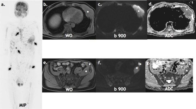
Figure 2.
Staging WB-MRI: A 65-year-old with non-secretory myeloma staging WB-MRI. The inverse grey scale b900 MIP (a), axial water only DIXON (b, c), axial b900 (d, e) and axial ADC (f, g) show multiple active lesions (arrows & asterisks). ADC, apparent diffusion coefficient; MIP, maximum intensity projection.

