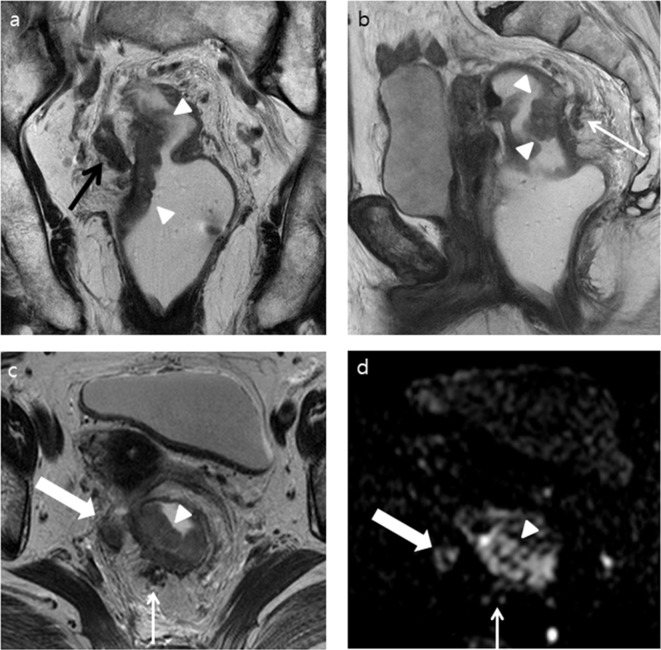
Figure 2.
A 81-year-old female with a pathologically confirmed rectal adenocarcinoma (pT3N2bM0) and positive EMVI. Both readers increased their confidence score for EMVI from 3 to 4 after adding DWI to T 2WI. (a) T 2WI coronal and (b) sagittal images show an ulcerofungating mass (white arrow heads) arising from the right posterolateral wall of the proximal rectum. Tumor invasion of the right mesorectal fascia is obvious (black arrow). Dilated peritumoral venous structure filled with tumor signal, located posterior to the tumor (white arrow), is also noted. (c) T2W axial image shows tumor (white arrow head) extending from the rectal muscularis propria to the neighboring serpiginous venous structure in the 7 o’clock direction (thin white arrow). The tumor penetrates into the right mesorectal fascia (thick white arrow) (d) DWI (b = 1000 s mm–2) shows a high signal intensity in the tumor (white arrow head) and in the presumed EMVI in the 7 o’clock direction (thin white arrow). An amorphous intermediate signal-intensity lesion corresponding to the tumor invading the right mesorectal fascia in the 9 o’clock direction (thick white arrow) is also noted. DWI, diffusion-weighted imaging; EMVI, extramural venous invasion; T 2WI, T 2 weighted imaging.

