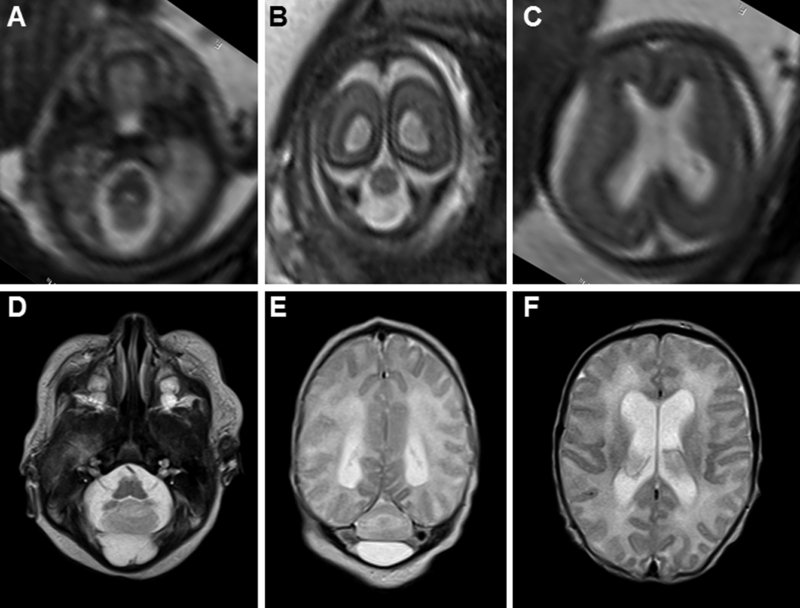
FIGURE 2.

Pre- and post-natal brain MRI features of RES. (A–C) Fetal MRI at 21 and 4/7 weeks gestational age demonstrates severe cerebellar hypoplasia (A and B) and mild ventriculomegaly (C). (D-F) Neonatal MRI of the same patient demonstrates severe cerebellar hypoplasia (D and E) and mild ventriculomegaly (F) in the same patient.
