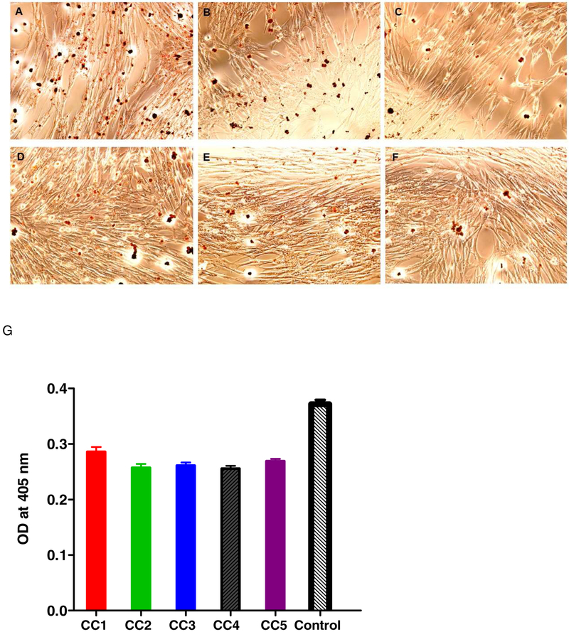
Figure 4.
LD staining using Oil Red O (ORO). ORO staining protocol described under Materials and Methods was followed. The paraformaldehyde (4%)-fixed cells were stained with ORO, followed by DAPI, and were visualized by fluorescent microscopy. (A) Control oleic acid-treated cells; (B-F): 5 μM curcumioids (CC1- CC5)-treated cells; (G) Quantification of LD after extraction by absorbance at 405 nm.

