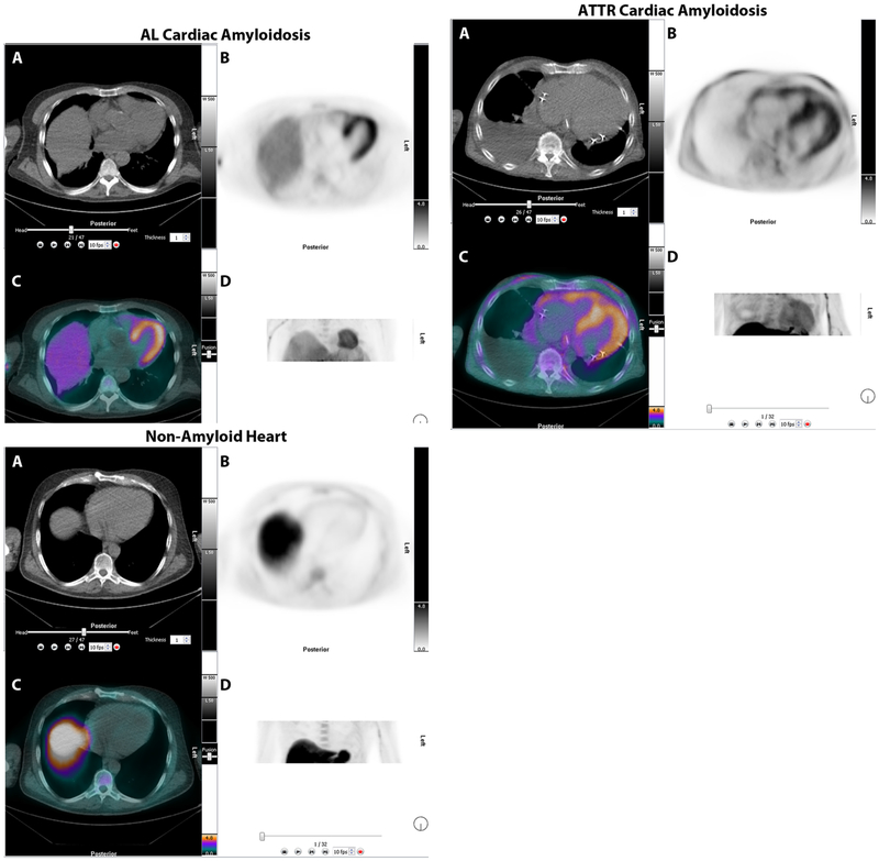
Figure 11.
18F-florbetapir PET/CT imaging in patients with AL, ATTR, and no-amyloidosis. Images are displayed as axial CT transmission (A), axial emission (B), fused transmission/emission (C), and a maximum intensity projection image (D). Both AL (top left), ATTR (top right) images show intense 18F-florbetapir uptake in the left ventricle. The patient without amyloidosis, volunteer, (bottom), showed no myocardial uptake of 18F-florbetapir.

