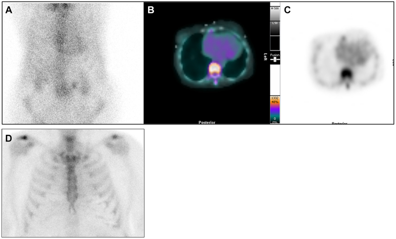
Figure 4.
99mTc-PYP on the 1-hour planar chest images and corresponding cardiac SPECT images. 99mTc-PYP with Grade 2 cardiac uptake on the planar chest images (A), which was confirmed as blood pool activity on SPECT/CT fusion images (B), and SPECT images (C). These are 1-hour images, and low rib uptake is noted. In contrast, Panel D shows 3-hour planar chest images from another patient showing Grade 0 (no) cardiac uptake and expected rib uptake.

