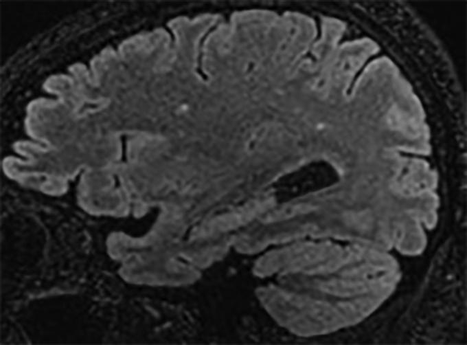
Figure 1.
A representative parasagittal volumetric T2 fluid-attenuated inversion recovery image of the brain demonstrating a few subcentimeter foci of signal abnormality confined to the white matter of the cerebral hemispheres. This is a nonspecific pattern that may be seen as a result of infection and inflammation (as well as prior trauma, migraine-related change, and primary demyelination). There is no evidence of meningitis.

