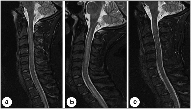
Figure 2.
Three midline sagittal short tau inversion recovery images of the cervical spine obtained throughout the clinical course of this patient. (a) At presentation, the image demonstrates longitudinally extensive signal abnormality in the spinal cord extending from the cervical-medullary junction into the thoracic spinal cord (not completely visualized). (b) Three days later, the cord became more edematous, resulting in further narrowing of the subarachnoid space, and signal abnormality became more evident in the brainstem, particularly the medulla. (c) Seventeen days after presentation, following corticosteroid and plasmapheresis therapy, and correlating with a profound clinical improvement, the image demonstrates nearly completely resolved signal abnormality and resolved edema in the brainstem and spinal cord.

