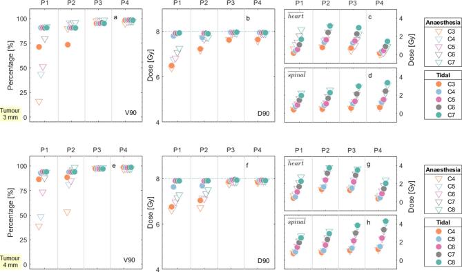
Figure 4.
DVH metrics for the four different tumour positions, represented by P1, P2, P3 and P4 at the abscissa axis, for the (a, b, c and d) 3 mm tumour and the e, f, g and h) 4-mm tumour. The circles and the triangles depict the anaesthesia and the tidal curves, respectively. The shades (or colours for the online publication) illustrate the different collimator sizes, ranging from 3 mm (C3) to 8 mm (C8). (a) and (e) show the V90 of the tumour, (b, f) the D90 of the tumour, (c, g) the mean dose to the heart and d,h) the mean dose to the spinal cord. The dotted line on (b, f) indicate the prescription dose of 8 Gy. DVH, dose-volume histogram.

