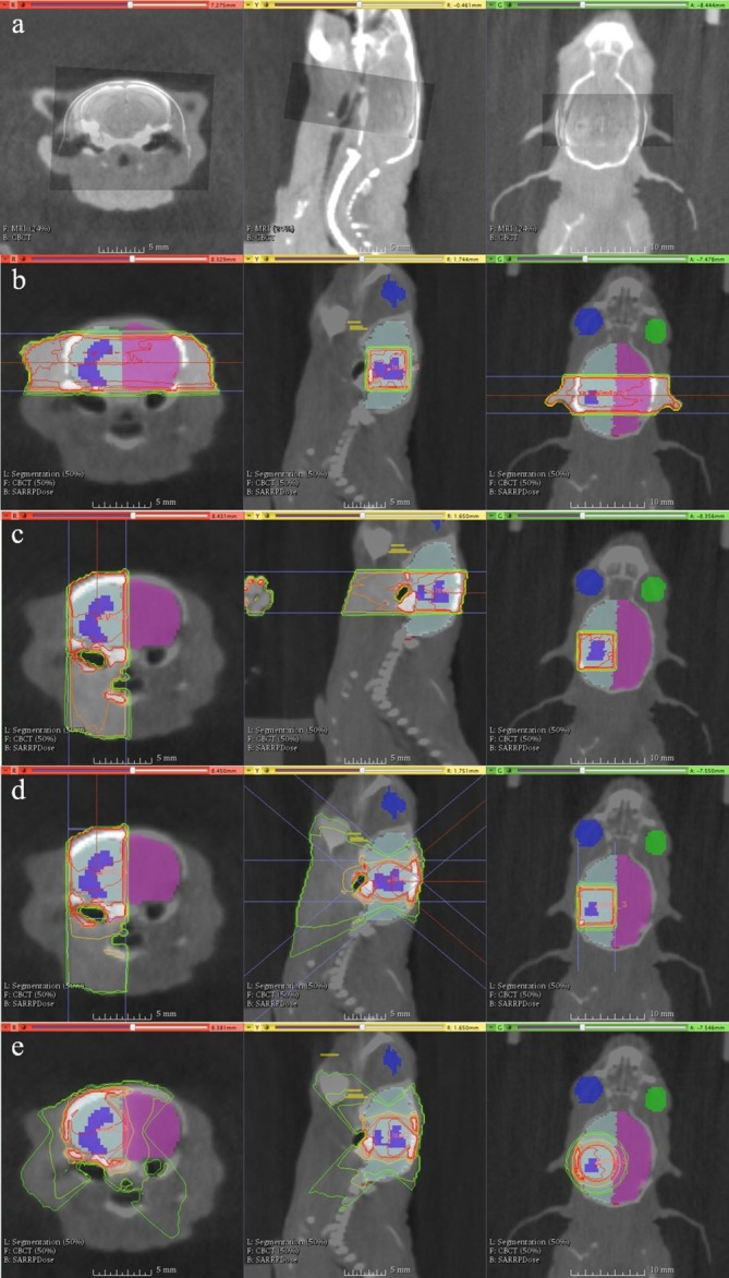
Figure 1.

Contoured colours: tumour (purple), ispilateral brain (light blue), contralateral brain (magenta), left eye (blue), right eye (green), mouth (yellow, largely out of viewing plane), brainstem (not shown). The lower left legends describe the current datasets in view in Muriplan, which can currently only show a background and foreground dataset, alongside contours.(a) MRI-CBCT fusion for target volume delineation. (b) Parallel opposed pair: two opposed lateral beams positioned so that the central axis passes through the centre of the tumour. (c) Single beam – single hemisphere: a single beam aimed “top-down” so that the central axis passed through the centre of the tumour, thus sparing the contralateral cerebral hemisphere.(d) Single plane arcs (“TopArc”) – single hemisphere: an arc that travels from gantry 40 to 0 degrees at couch 90, and then from 0 to 40 degrees at couch −90 to form an arc over the tumour from above, with the aim of sparing the contralateral hemisphere of the brain while improving tumour conformity.(e) Couch rotation arcs (“ArcXX”): fixed gantry set up with a complete couch rotation designed to create a spherical type dose distribution. Gantry angles, XX, of 30, 45 and 60 degrees were used to modulate the sphericity of the distribution. In the example shown the gantry angle is 45 degrees. CBCT, cone beam CT.
